Bursitis: what is it and which parts of the body are most at risk
Swelling, pain, difficulty in moving the joint: these could be signs of bursitis, a very common inflammation which appears mainly in the elbow, knee, shoulder, hip or foot. In this article, we illustrate the most frequent types of bursitis and understand how to intervene and what the recovery times are for returning to full joint mobility and functionality.
What is bursitis and what are the main causes
Bursitis is the inflammation of the bursa, a small, flat sac filled with liquid located between the muscles, tendons and bones. The function of these bursae is fundamental for reducing friction and protecting the joint structures from everyday stresses.
In general, there are two types of bursitis, inflammatory and haemorrhagic. The causes of the first type relate to excessive rubbing or overload (very common among sportspersons, for example), repeated micro-traumas over time and incorrect postures, tendon problems such as inflammation or tissue degeneration, or by bacteria following skin lesions. Haemorrhagic bursitis, on the other hand, is caused by an acute trauma, which in this case causes blood outpouring into the bursa.
Symptoms
The symptoms of bursitis obviously depend upon the position of the inflamed bursa. Generally, however, there is acute pain and discomfort in the affected area, often accompanied by visible and/or palpable swelling associated in some cases with hot spots due to an increased body temperature in the inflamed area.
The most common types
The areas most commonly affected by bursitis are:
- The shoulder (subacromial bursitis)
- The elbow (olecranon bursitis)
- The knee (prepatellar bursitis)
- The hip (trochanteric bursitis)
- The internal part of the knee (goosefoot bursitis)
- The Achilles tendon (Achilles tendon bursitis)
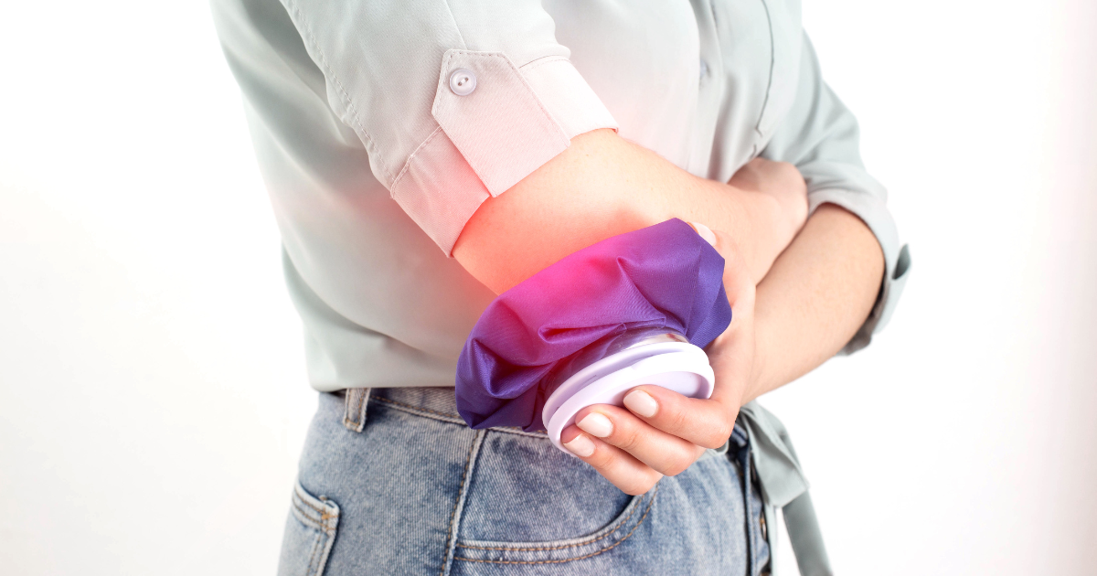
Bursitis of the elbow or olecranon
The olecranon bursa, located between the olecranon of the ulna and the skin, can become inflamed for various reasons, particularly acute or repeated traumas. This type of bursitis causes swelling at the rear of the elbow, in some cases even without pain, but which can arise or be exacerbated simply by pressing on the area, such as resting the elbow on a table.
Subacromial bursitis of the shoulder
There are a number of bursae in the shoulder, but the one that most frequently becomes inflamed is the subacromial bursa. This bursa is located between the acromion of the shoulder blade and the humerus, and helps to reduce the friction between these two bones and the rotator cuff tendons. Typically, it causes pain in the front and side of the shoulder. Repeated movements of the arms above the head (so-called overhead activities in sporting jargon) are painful and the symptoms often worsen during the night.
Prepatellar bursitis of the knee
Also known as “housemaid’s knee”, this inflammation is caused by an acute trauma such as falling on the knee, repeated micro-traumas such as working frequently on your knees or other causes such as an infection. The knee also has several bursae, but the most affected are the prepatellar (between the patella and the quadriceps tendon) or the infrapatellar bursa (between the tibia and the patellar tendon). There is usually pain in the front of the knee, associated with swelling and limited movement.
Trochanteric bursitis
In the case of trochanteric bursitis, the two most frequently affected bursae are those in the gluteus medius and gluteus maximus, localised between the greater trochanter of the femur and the tendons of the related muscles. The main symptom is pain, localised in the side of the hip, above all when climbing the stairs or sleeping on one side. It is however more difficult to feel swelling or heat. These bursae become inflamed as a result of direct traumas in the area, such as falling on one side, or chronic overloads due, for example, to postural alterations or lumbar or pelvic dysfunctions.
Goosefoot bursitis
The goosefoot, or pes anserinus, inserts onto the sartorius, gracilis and semitendinosus muscles conjoined on the proximal and medial part of the tibia. The bursa lies precisely between the insertion of these tendons and the tibia, to reduce friction. Also in this case, the symptoms appear around the knee, but in contrast to prepatellar bursitis, with goose foot bursitis the pain lies in the front and middle of the knee and is associated to swelling and heat in the area.
Achilles tendon bursitis
Two bursae are affected by this condition: the first lies between the Achilles tendon and the skin, behind the heel (posterior bursitis) while the second is localised between the Achilles tendon and the calcaneus (anterior bursitis). In both cases, the main symptom is pain in the rear of the heel, but with posterior bursitis, which has a more superficial anatomical position, the skin may appear reddened and swollen.
What to do in the case of bursitis: diagnosis and treatment
Considering that the symptoms are also common to other joint conditions, the first step is to obtain a differential diagnosis from a specialist Physiatrist or Orthopaedic doctor. The diagnosis is based exclusively on a physical examination or instrumental examinations such as an ultrasound scan or magnetic resonance.
Having confirmed the diagnosis and excluded other joint problems, the treatment of bursitis caused by a trauma or repeated movements (so without infection) includes:
- Resting the affected joint;
- Ice;
- Instrumental therapy such as LASER, TECAR or Ultrasound;
- Anti-inflammatory drugs;
- Therapeutic exercises, planned according to the perceived pain;
In the case of bursitis caused by an infection, the physician will prescribe the most suitable antibiotics and if required may aspirate the liquid from the bursa. In these cases, it is absolutely vital to avoid cortisone infiltrations, as this may worsen the situation.
Recovery times and the importance of physiotherapy
Recovery times depend upon the site and extent of the inflammation and may therefore vary from a few days to several weeks.
During acute phases, often a period of rest and anti-inflammatory drugs are sufficient, but with chronic bursitis physiotherapy is fundamental for understanding the actual cause of the problem. In fact, chronic bursitis is often caused by postural alterations or dysfunctions that lead to continuous overload of the affected joint, preventing the inflammation from healing. A correct physiotherapy, osteopathic or postural assessment will be able to analyse the main cause of the problem, then drafting an effective, targeted recovery plan aiming to completely heal the bursitis.


