Tendinitis: how it develops and how to treat tendon inflammation
Tendinitis is an inflammation caused by overload, affecting not only athletes and sports men and women, but also those who perform continuous and repetitive movements or those suffering from postural problems. This article will look at the causes, symptoms and the tendons most susceptible to this type of injury.
Tendons are fibrous structures that bind muscles to bones or other insertional structures. Tendons are made up of two substances: one highly resistant, known as Collagen, and a more flexible one known as Elastine. Their main function is to transmit the strength of the muscle to the structures connected to it, so that the joints move correctly.
Given their function, tendons are found in numerous parts of the body, but those most susceptible to injury and inflammation are without a doubt the tendons of the shoulder, elbow, hand, knee and the well-known Achilles tendon, i.e. the tendon connecting the posterior muscles of the calf to the foot.
Tendon inflammation: symptoms and causes of Tendinitis
Tendinitis is essentially the inflammation of a tendon. The main symptom of tendinitis is pain, which tends to worsen with movement or under pressure. Other symptoms related to inflammation of the tendon are heat, swelling or reddening.
In acute cases of tendinitis, the cause is often traumatic, such as a sprain or excessive load during training. Otherwise, in the case of chronic tendinitis, we need to investigate the cause by repetitive and continuous movements over time – for example due to work tasks – or possible postural problems.
Which tendons are most susceptible to inflammation?
Rotator cuff tendinitis
This is an inflammation of the shoulder tendons, which therefore affects the main stabilisers of the glenohumeral (or scapula-humeral) joint, i.e. the muscles that make up the rotator cuff. The tendons most susceptible to inflammation are those of the supraspinatus muscle, often due to a condition known as Subacromial Impingement, where the tendon in question is repeatedly crushed between the humeral head and the acromion (anatomical part of the scapula) while lifting and lowering the arm.
Shoulder tendinitis is one of the most common problems related to the scapula-humeral joint, and manifests as a sharp pain in the shoulder, above all overnight. If you have recently had pain in the shoulder and you think it may be due to a tendon injury, have a look at the tips from our physiotherapists to learn about what to do in the case of inflammation of the rotator cuff.
Elbow tendinitis
When we talk of tendon problems related to the elbow, this mainly concerns lateral epicondylitis (commonly known as Tennis elbow), which causes pain on the outer side of the elbow, or medial epicondylitis (so-called golfer’s elbow), if the pain is located on the inner side of the elbow.
While these are two different disorders, both cases are caused by an overload on the tendon, in which symptoms arise when heavy objects are lifted or when the hand is squeezed. In the most serious cases, elbow pain persists also at rest or after minor exertion, such as opening a bottle of water, and often extends to the forearm, wrist and occasionally to the fingers.
Knee tendinitis
The most common cases of knee tendon inflammation affect:
- Patellar tendon or patellar ligament: the central part of the knee, connecting the patella to the tibia. Patellar tendinitis is caused by functional overload, mainly due to repeated jumping actions – indeed it is commonly known as Jumper’s knee and mainly affect sports men and women.
- Iliotibial band: in other words, the external part of the knee In this case inflammation impacts the tissue that on the outer side of the thigh extends down to below the knee. The so-called “iliotibial band syndrome” can occur after movements repeated straightening and bending the knee over a prolonged period, such as during running or cycling.
- Pes anserine tendinitis (Goose foot tendinitis): the tendons that form the goose foot connect the sartorious, the gracilis, and the semitendinosus muscles on the inner part of the tibia, thus forming the medial section of the knee, responsible for bending and internal rotation of the joint. Constant stress causes inflammation, which manifests with sharp stabbing pain below the knee, often accompanied by swelling, both of which persist even at rest, especially at night.
When the knee tendons are inflamed, pain can occur in many of the movements that involve the lower limbs, such as running, jumping, going up and down stairs and simple daily chores, which means particularly unpleasant symptoms if not treated promptly.
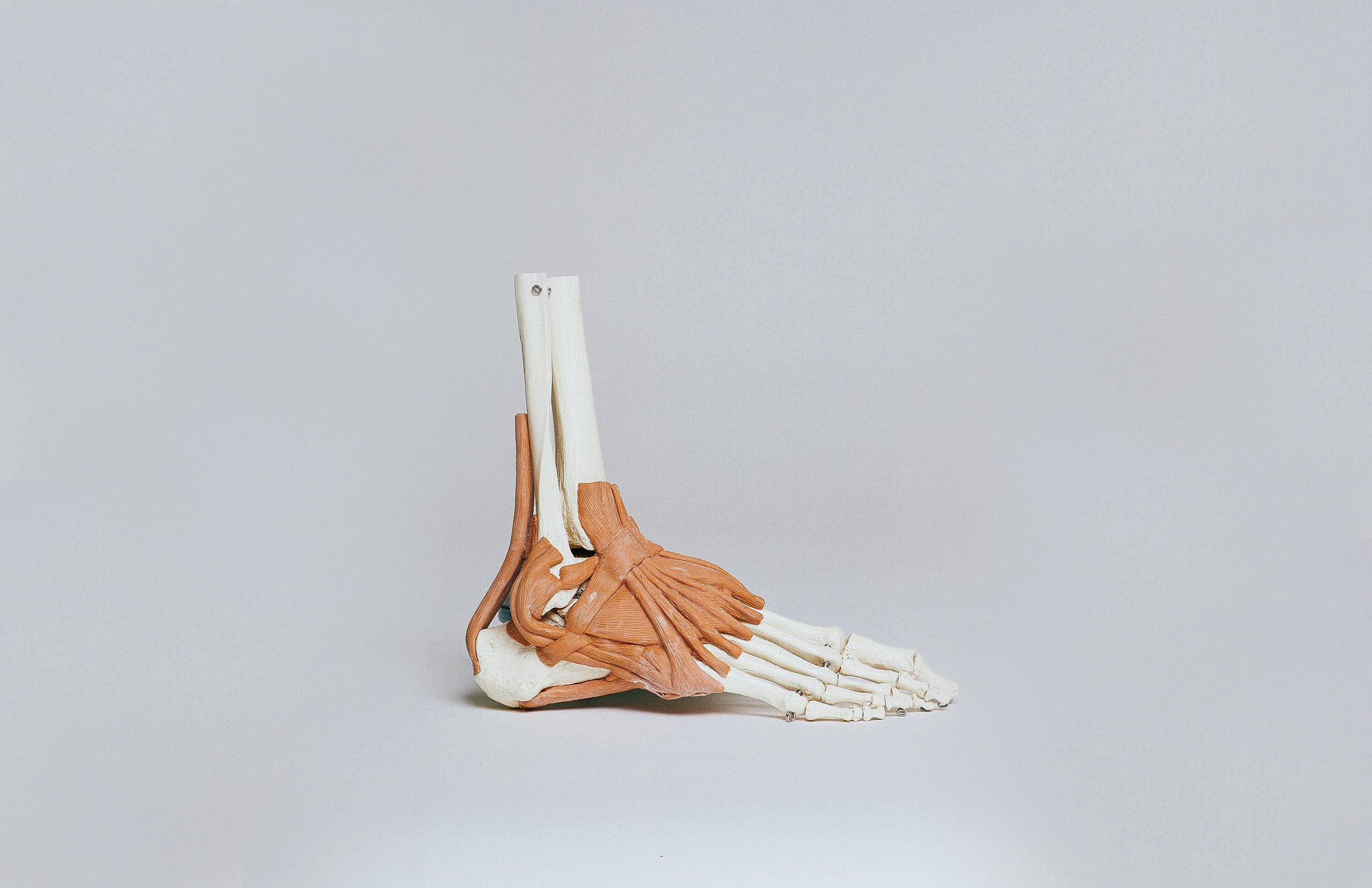
Achilles tendinitis
The Achilles tendon is a highly resistant structure connecting the triceps surae muscle to the calcaneus (heel bone). For various reasons, including overload, traumatic injuries, altered arch support or rigidity of the muscles in the posterior chain, the tendon may become inflamed.
Pain is normally located in the lower area near the heel bone during activities such as walking, running and jumping, but also when pressing on the back of the ankle. Sharp, shooting pain is often accompanied by a loss in muscle strength and are particularly acute in the morning after waking when taking the first steps, to then subside gradually over the day. As well as pain in the foot and ankle, inflammation of the Achilles tendon can lead to swelling, stiffness and in some cases, the appearance of erythema.
Effective treatments for tendinitis
The duration of acute tendinitis can vary greatly, and recovery times are very subjective. It may take just a few days, or even a few weeks for complete recovery.
Acute tendinitis is initially treated with rest, the repeated application of ice and possibly the use of anti-inflammatory medication. Instrument-assisted therapies can also help, such as Laser or Tecar in athermal mode. This initial therapy is usually integrated with motor rehabilitation of the muscle and tendons with specific exercises.
In the case of chronic tendinopathy, undoubtedly the most complex to treat, it is also essential to identify the cause of the problem to then establish the correct rehabilitation programme together with a physiotherapist. In these cases, before starting out on the recovery therapy, some diagnostic examinations are useful, such as an ultrasound or magnetic resonance image, to check the condition of the tendon and the possible presence of calcification. Only in this case, it will be the doctor’s decision whether surgery is needed on the injury.
 The most common injuries in the mountains: how to prevent them and how to recover quickly
 Post-workout muscle recovery and DOMS prevention: tips, effective exercises and management strategies
 Meniscus and cartilage wear: causes, treatments and useful exercises
 Knee pain: causes, symptoms and remedies


