
Dislocation: what it is and how to treat it to reduce recovery times
A dislocation is one of the most frequent and painful joint traumas, not to be confused with a sprain. A dislocation is in fact a traumatic displacement of the joint, which occurs when the two bone heads in the articulation shift permanently beyond the physiological articular range. When there is a total separation of these joint surfaces, we talk of complete dislocation, while if the dislocation is partial we talk of subluxation.
Which joints are most at risk of dislocation, and how can we intervene?
In the vast majority of cases, the dislocation is caused by a trauma, which can occur in many different ways, such as during contact sports, a car accident or a simple fall. Some joints are certainly more at risk of this type of trauma than others, such as the shoulder, knee or hip.
The most frequent types of dislocation are described below, with indications of first aid measures as well as post-trauma recovery:
Dislocated shoulder
A dislocated shoulder is certainly the most frequent, counting for around 50% of cases. The triggering trauma is often a fall, with the weight on the shoulder joint or on a straight arm. In this type of trauma, the separation occurs between the shoulder blade and the head of the humerus, which comes completely out of the glenoid cavity. In most cases, a dislocated shoulder occurs at the front, when the humeral head comes out of its seat anteriorly.
Unless the dislocation returns spontaneously into its seat, it is fundamental to go immediately to the accident and emergency department, where expert doctors can use specific manoeuvres to put the joint back in place. Never ask a friend to reduce the shoulder into position!
In fact, in some cases, the dislocation may be associated to a fracture, or vascular or nerve damage, and these could worsen when not managed by experts, causing even permanent damage.
If there is no other associated damage, once the joint has been reduced a sling should be worn for a few weeks, to give the tissues time to heal. Thereafter, patients should undergo physiotherapy, with an expert physiotherapist who can define a recovery programme to strengthen the joint, the muscles and proprioceptive training.
In some cases, even after rehabilitation, the dislocation may leave the shoulder joint unstable and therefore at risk of subsequent dislocation, even in the event of mild traumas. In these cases, unfortunately the only solution is surgery, aiming to stabilise the joint structure.
Hip dislocation
In hip dislocations, the bones involved are the head of the femur, which separated from the acetabulum. Usually, in healthy persons the cause is a severe trauma, such as a head-on car accident or a fall from a height, for this reason this type of dislocation is often also associated to one or more fractures.
Depending on the severity of the dislocation and any associated damage, the emergency doctor will decide whether to proceed with manual reduction of surgery. In both cases, physiotherapy is fundamental for rehabilitation.
In the case of the hip, in addition to traumatic dislocation there is also congenital dislocation, known as congenital dislocation of the hip. This is a malformation which gradually leads to the dislocation of the femural head from the acetabulum. This defect is due to an abnormal development of the hip joint before birth, which unfortunately leads to walking difficulties. This is why all children, between the second and third month of life, undergo a specific ultrasound scan, to be able to promptly diagnose any congenital dysplasia and intervene using specific devices depending on the severity.
Elbow dislocation
Elbow dislocations are also quite frequent; in this case, there is a loss of contact between the humerus (arm bone) and the two bones in the forearm, the ulna and the radius. Elbow dislocations are usually due to a fall with the arm extended, bike or motorbike accidents or head-on car accidents, when the victim instinctively extends the arms forwards as if to defend themselves. In children, the common causes of elbow dislocation are sudden tugging of the wrist or lifting them up by the wrists.
Also in this case, elbow dislocations can be associated to one or more fractures, which must be assessed with an x-ray at the accident and emergency department. Depending on the clinical situation, the arm may be immobilised with a conventional plaster cast or a sling, or surgery.
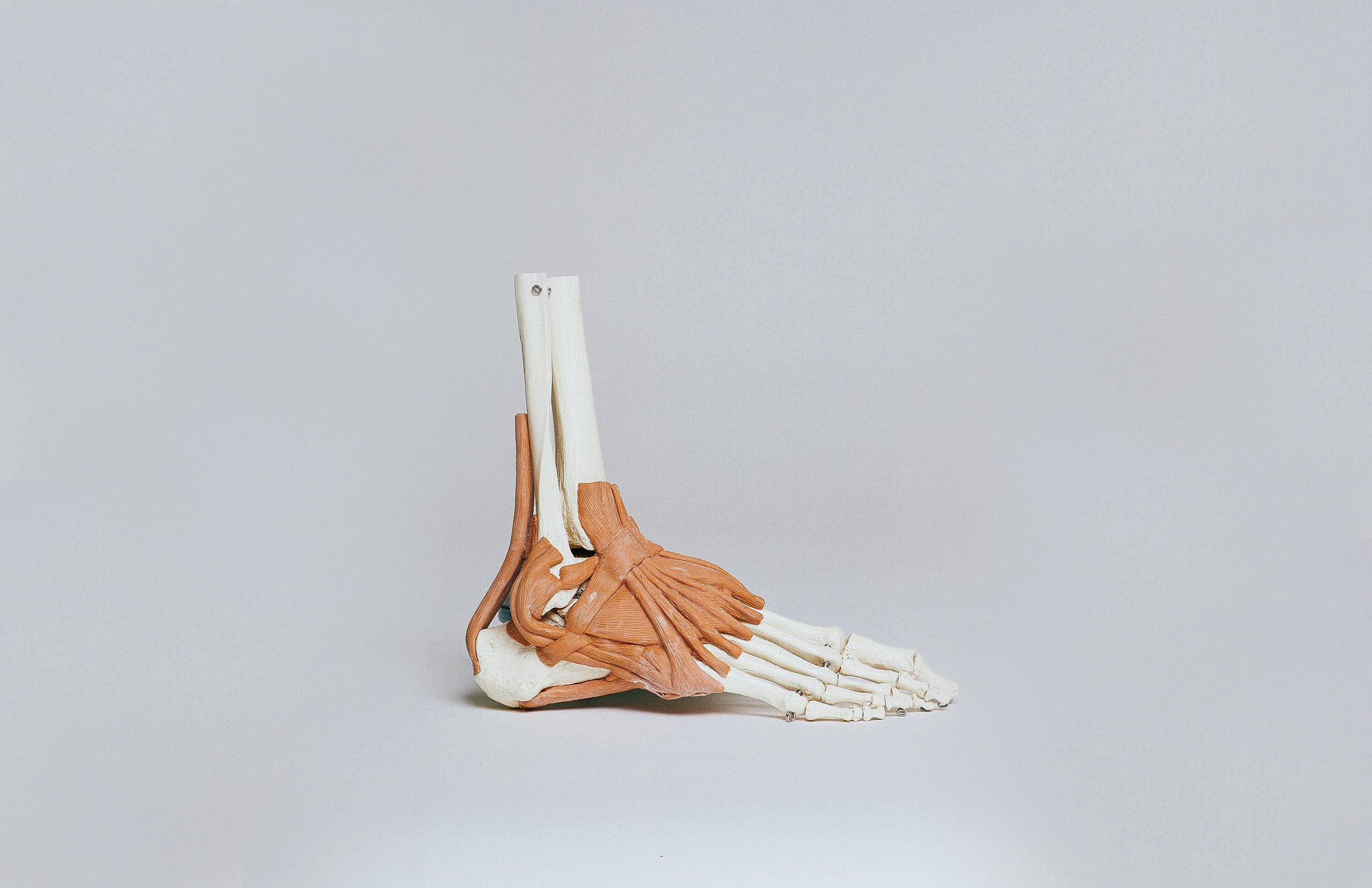
Patellar dislocation
Dislocation of the knee is when the patella slips completely out of the femural trochlea (the seat in the final part of the femur). Patellar dislocation occurs mainly as a result of trauma, but in some cases it may be related to congenital ligamentous laxity, the excessive elasticity of the ligaments.
In the case of the knee, with a dislocation caused by trauma or a fall, there is pain, swelling, difficulty walking and the feeling that there is “something out of place” in the joint. Very often the dislocation reduces on its own after a trauma, but a visit to the A&E is always advisable, for an x-ray to exclude any fractures. In most cases, the treatment is conservative, using a knee brace for a few weeks, followed by a cycle of rehabilitation aiming to recover the joint movement in the knee, strengthen the muscles and proprioceptive training.
Dislocation of the acromioclavicular joint in the shoulder
This is quite a frequent shoulder injury, both complete and partial (subluxation), affecting the acromioclavicular joint, which is the end of the lateral clavicle and the shoulder blade. In contrast to humeral dislocations, in this case the joint is held in place by some ligaments which are strained or torn during the dislocation. Very often this dislocation is caused by falling on the shoulder or following a contact-sports trauma (rugby, football, etc.).
Clinical examinations will show a clear swelling and bruising in the joint, with shooting pains when touching or moving the shoulder. Another characteristic sign of acromioclavicular dislocation is what is known as “piano key”: pressing on the joint, the doctor or physiotherapist lowers it, returning the joint to its seat, but the joint rises again once the pressure is removed. An x-ray is usually done to confirm the diagnosis.
Depending on the severity of the dislocation, and how may and which ligaments were torn during the trauma, the orthopaedic specialist may decide on conservative treatment or surgery. In the first case, a sling is worn for several weeks, followed by physiotherapy. In the second, surgery is done to stabilise the joint, followed by rehabilitation.
Other less frequent types of dislocation
Other less frequent types include dislocation of the temporomandibular joint, dislocation of the coccyx and the sternoclavicular joint.
- Dislocation of the TMJ (temporomandibular joint): this is the joint between the jawbone and the temporal bone, which allows the mouth to move. In the event of a dislocation, the joint remains out of place after the person has opened the mouth wide, and cannot then close it. Often it is reduced on its own, but it may cause pain and inflammation for several weeks. To understand the causes of the dislocation, the joint must be checked by a dentist or gnathologist.
- Dislocation of the coccyx: the dislocation of the coccyx always occurs due to a direct trauma in the sacral area, such as a hard fall on the back or during natural childbirth. The symptoms are pain in the lower region of the sacrum, back pain and difficulty sitting. The treatment includes pain drugs, with manipulation by a physiotherapist or osteopath, aiming to reduce the muscle and ligament strain and help the coccyx to return to its physiological position.
- Dislocation of the sternoclavicular joint: dislocation of the sternoclavicular joint is that of the sternum and the clavicle bone. In the event of dislocation, which is very rare, the clavicle leaves its seat, causing an anterior or posterior dislocation. Although anterior dislocations are easy to resolve, (less frequent) posterior dislocations can be dangerous and must be identified and treated rapidly, often surgically. The most frequent cause is a shoulder trauma, which then affects this joint.
Depending on the severity of the dislocation, once assessed with an x-ray, the orthopaedic specialist may opt for conservative treatment or surgery in order to stabilise the joint.
Risk factors
As in the vast majority of cases dislocation is a traumatic event, it cannot be prevented. The only advice we can give is always to maintain a good level of muscle tone and suitable proprioception, which we can achieve with a varied, constant physical training involving the whole body.
The only real risk factor is congenital ligamentous laxity, which causes the ligaments to be overly elastic, making the joints less stable. Also in this case, we can increase muscle tone and joint proprioception through appropriate physical exercise, in order to compensate the excessive elasticity.
What to do and, especially, what NOT to do in the event of a dislocation
In the event of a traumatic dislocation, go as soon as possible to the A&E department, both to reduce the joint and assess any damage to the bones, ligaments and surrounding nerves. Caution! Never attempt to reduce the joint on your own or with the help of anyone who is not an expert, as this could cause irreversible damage.
Only an x-ray or CAT scan can help to assess the state of the dislocation and any fractures. Depending on the outcome of the diagnostic tests, the doctor can decide whether to reduce the dislocation manually or arrange for surgery. In the first case, a sling must be worn for a few weeks, followed by rehabilitation with a physiotherapist to recover joint movement and help muscular and proprioceptive strengthening. In the case of surgery, on the other hand, the rehabilitation programme is assessed according to the severity of the case.
Recovery times
Recovery times vary according to the site of the dislocation, but generally after a few weeks of rest, with the aid of a sling, a rehabilitation programme lasting an average of 2 months enables a return to normal everyday activities, while the return to competitive sports takes up to 4-6 months in the case of a dislocated shoulder.
In fact, with a dislocated shoulder, joint instability may last, causing chronic pain and repeated dislocations. In these cases, the orthopaedic specialist and physiotherapist have to decide whether to continue with rehabilitation or opt for surgery to stabilise the joint.
Both with conservative rehabilitation and after surgery, return to sports activity is usually possible with the authorisation of the orthopaedic specialist, who assesses the suitable time frame.
 The most common injuries in the mountains: how to prevent them and how to recover quickly
 Tendinitis: how it develops and how to treat tendon inflammation
 Post-workout muscle recovery and DOMS prevention: tips, effective exercises and management strategies
 Meniscus and cartilage wear: causes, treatments and useful exercises


